


12 Mar Neuroradiology for Traumatic Brain Injury Diagnosis
By visualizing the internal structures of the head and spine, neuroradiology aids in the precise diagnosis of various neurological conditions. Medical professionals rely on these detailed images to accurately diagnose and assess conditions or injuries. This field provides the foundational information necessary for medical teams to develop appropriate, individualized care plans. A traumatic brain injury (TBI) occurs when a sudden physical force impacts the head and disrupts normal brain function. Here’s information on neuroradiology services for traumatic brain injuries and other health conditions:
What Causes Brain Injuries?
Brain injuries often result from forceful impacts or sudden changes in momentum. Some common causes include motor vehicle accidents, accidental falls, sports-related collisions, and physical assaults. Rapid acceleration and deceleration can also cause the brain to collide with the inside of the skull. When these events occur, individuals may experience a range of physical, cognitive, and sensory symptoms. Neuroradiology utilizes specialized imaging techniques, allowing specialists to identify and evaluate these neurological injuries.
Traumatic brain injuries range from mild concussions that cause temporary dysfunction to severe structural damage that requires extensive evaluation. Some individuals might experience changes in sleep patterns or noticeable shifts in mood following the incident. Physical signs frequently involve persistent headaches, localized pain, dizziness, fatigue, and sensitivity to bright lights. Some cognitive indicators of a TBI may include:
- Memory Loss
- Acute Confusion
- Concentration Difficulties
Neuroradiology is a branch of radiology that diagnoses abnormalities of the central and peripheral nervous systems, offering safe ways to visualize the brain, spine, and surrounding anatomical structures. This field utilizes advanced neuroimaging technologies to capture cross-sectional pictures of the nervous system. Medical providers use these accurate radiological reports to understand the specific physical nature of an individual’s injury. These imaging scans enable neuroradiologists to identify internal bleeding, tissue swelling, skull fractures, and microscopic damage.
What Diagnostic Scans Are for TBIs?
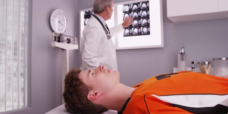
When a person has a potential head injury, medical teams rely on specific imaging tools to safely evaluate the damage. Computed tomography (CT) scans are typically the first assessment recommended for acute head trauma. A CT scan uses rotating X-ray machines and computer processing to create detailed, cross-sectional images of the skull and brain. This scan quickly reveals bone fractures, active hemorrhaging, and large hematomas. The speed of a CT scan makes it highly beneficial during emergency medical situations.
Magnetic resonance imaging (MRI) is another powerful diagnostic tool for neurological evaluations. An MRI uses strong magnetic fields and radio waves to generate highly detailed images of the brain’s soft tissues. A CT scan is generally faster, but an MRI provides greater detail for detecting subtle damage and micro-hemorrhages. CT scans identify immediate structural threats, while MRIs detect subtle abnormalities in the brain tissue. These scans benefit TBI diagnosis by pinpointing the exact location of the injury, allowing physicians to guide necessary medical interventions and monitor healing.
What Are Other Neuroradiology Scans?
Neuroradiology encompasses several other specialized imaging tests, in addition to CT and MRI, to evaluate the nervous system. Cerebral angiography focuses specifically on analyzing the blood vessels within the brain and neck. During this imaging procedure, a specialist injects a contrast dye into a major artery and takes a series of X-rays. This scan highlights factors that may be associated with severe head trauma, such as:
- Blockages
- Vascular Damage
- Aneurysms
Myelography is another specialized imaging procedure that examines the spinal canal, spinal cord, and nerve roots. A physician carefully injects a contrast material into the spinal fluid space before taking continuous X-ray images. Cerebral angiography visualizes the vascular network, and myelography highlights issues within the spinal column. These scans enable a comprehensive evaluation of the central nervous system for other neurological concerns besides TBIs.
Contact a Radiology Specialist Today
Neuroradiology provides the clear, precise visual information needed to understand the extent of head trauma. Getting a prompt diagnosis is a fundamental step in managing any neurological condition, as brain injuries can affect many aspects of health. Contact a radiology specialist today to learn more about available imaging procedures, schedule an appointment, and discuss your testing options.
- How to Create Web Applications Using OneFramework
- Does SouthSlopeNews.com Cover Local Events? A Complete Guide
- How Does PresidentialSummary.com Work? A Complete Guide for Readers
- How Often Does Manga Buddy Update? Everything Readers Need to Know
- Can I Read Manga Online on Omega Scans? A Complete Guide for Manga Fans
No Comments